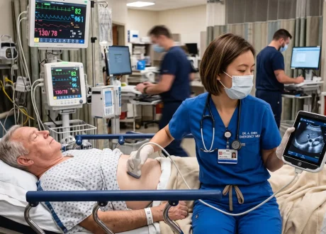

***It’s no secret that ultrasounds are not my biggest strength. When I run into trouble with complicated Echos I turn to my friend and echocardiography guru Aram, who is part of the team who writes the “The Echo Journal” This week, we dive into a complicated patient with an interesting finding on echo!***
“It’s happening again, Doctor. I brought him back to see you. You’re the only one I trust.”
As you walk into room 26 you see a familiar face. In the room is Mrs. Jackson, someone you remember quite well. 5 years ago, you diagnosed her father with pneumonia. He could barely breath. You treated his fever, started IV antibiotics and fluids, and intubated him when he could no longer breathe on his own. You talked Mrs. Jackson through every step, every decision, and explained your concern that he may not make it through the night, despite your best efforts. You know that her father was in the ICU, intubated, for 6 days and survived. And you haven’t seen him or Mrs. Jackson since they were discharged.
“It’s happening again, Doctor. I brought him back to see you. You’re the only one I trust. We moved away when Dad got better. Out of the city to a calmer life for him. But the fevers are back. And when he gets sick, we come back here. To see you. You remember he has that liver transplant and diabetes; we worry about him all the time. At the last hospital, no one could figure it out and we got scared. 3 days admitted and no answers. So, we left and came here. To see you.”
“Tell me what’s going on?” you ask her.
“Dad has had a fever for over 7 days. We thought it was nothing at first. But then it got worse. He has shaking spells now where he says he’s frozen cold. And he gets delirious sometimes too. So, we went to the local hospital. Thought it was the pneumonia back again, but the chest x-ray looked ok. So, who knows? And the fevers persist. The antibiotics aren’t helping. I’m very worried.
“Sir, how do you feel?” You ask Mrs. Jackson’s father.
“Tired and weak. I think I’m dying,” he tells you.
Your charge nurse, Shannon, walks in the room to help get the patient hooked up to the monitor and get some vitals. She also gets him hooked up to the ECG machine and hands you the completed ECG:

Rate: 130s
Rhythm: Sinus Tach
Axis: Normal
Intervals: Narrow QRS complexes, normal PR interval, QTc looks ok too
Morphology: Occasional PVCs (beats 3, 5, 7). There is also some concerning ST segment depression and T wave inversion in V3 – V5. I don’t love the way those T waves look at all.
Final Read: Sinus tach with concerning T waves in V3-V5.
“Doc, vitals coming at you,” Shannon says. “Heart rate is 137, blood pressure is 82/31, Resp rate 18, SpO2 90% on room air. Oh, temp is 103.5 and glucose normal. I’m going to activate him as a sepsis alert.”
“His fever is so high, he needs help, Doctor!” Mrs. Jackson says and looks at you with concern.
“I agree Ma’am. I’m very worried too. Mrs. Jackson, when you were at the last hospital, do you remember what work up was done and if they found anything?”
“Well, Doctor, they started off thinking it may be a virus, since his swab was positive for rhinovirus, but he just kept getting worse and worse. They put him on antibiotics just in case, even with the positive viral swab. By day 2 with no improvement despite IV fluids and IV antibiotics, they started looking deeper for a source of infection. The blood cultures finally grew out some kind of bacteria. And they said that he had a ‘weird’ pericardial effusion. They wanted to do a CT scan, but I said no. Send him over here. To see you. When they said no, I signed him out and drove him here myself.”
“Ok, thank you, that’s very helpful,” you tell Mrs. Jackson. “Let’s do a quick ultrasound of the heart to take a look at this effusion and see what the other hospital was seeing:”
This seems like an awfully complex ultrasound. Let me run it by Aram:
****
First Thoughts
The parasternal long axis (PLAX) view shows biventricular dysfunction right away. The short axis view (PSAX) at the aortic valve level is off-axis and technically challenging, with an anechoic structure adjacent to the right ventricle on the left side of the screen (Red arrow). Sliding to the LV papillary level, we confirm biventricular dysfunction from an oblique window. Moving further to the apex, echogenic material comes into view in or around the right ventricle.
The apical views were too limited to fully assess the RV free wall, so we lean on the subcostal window instead. It delivers, confirming a mildly echogenic space adjacent to the RV free wall.
So, what is going on here?
Final Read
There is a large pericardial effusion with fluid tracking adjacent to the right ventricle, best visualized in the subcostal window.
Although the RV appears compressed by the effusion, there is no significant respiratory variation in mitral or tricuspid inflow, and the IVC collapses normally: this argues against echocardiographic evidence of tamponade.
The LV ejection fraction is severely reduced at 20-25%, with multiple large wall motion abnormalities aslo present. Mild mitral regurgitation and mild aortic regurgitation are also present.
How Can We Tell the Difference Between Abscess and Effusion?
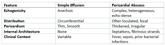
On 2D imaging, a pericardial abscess does not look like a typical effusion. Instead of a simple anechoic (black, fluid-filled) stripe, you are looking for a complex, echo-dense or heterogeneous collection, often with internal septations, fibrinous strands, and a thickened, shaggy-appearing pericardium. The collection may appear loculated rather than circumferential, meaning it is walled off in one area rather than surrounding the entire heart. This loculation is a key clue. Pus does not flow freely the way serous fluid does, so it tends to collect adjacent to a single chamber rather than layering posteriorly and inferiorly the way a simple effusion typically does.
In some cases, the abscess presents less like an effusion and more like a discrete mass: a walled-off, encapsulated collection compressing an adjacent chamber — which is why it’s occasionally mistaken for a pericardial cyst or tumor before the infectious etiology becomes clear.
Keep in mind TTE has real blind spots here, particularly for posterior loculations. If clinical suspicion is high but the transthoracic windows are inconclusive, TEE or CT should be pursued rather than assuming a negative TTE rules out an abscess.
Let’s sum it up with a nice table:
Thanks, Aram!
****
“Doc, what is that?” Shannon asks you, pulling you out of the US machine.
“That’s certainly concerning for a pericardial abscess!” you tell her. Let’s get him to CT emergently and I’ll give a call to interventional cardiology and CT surgery to give them a heads up. Mrs. Johnson, I think we may have found the source of your Dad’s fevers!”
Case wrap up:
Radiology calls you and asks you to take a look at the patient’s CT:
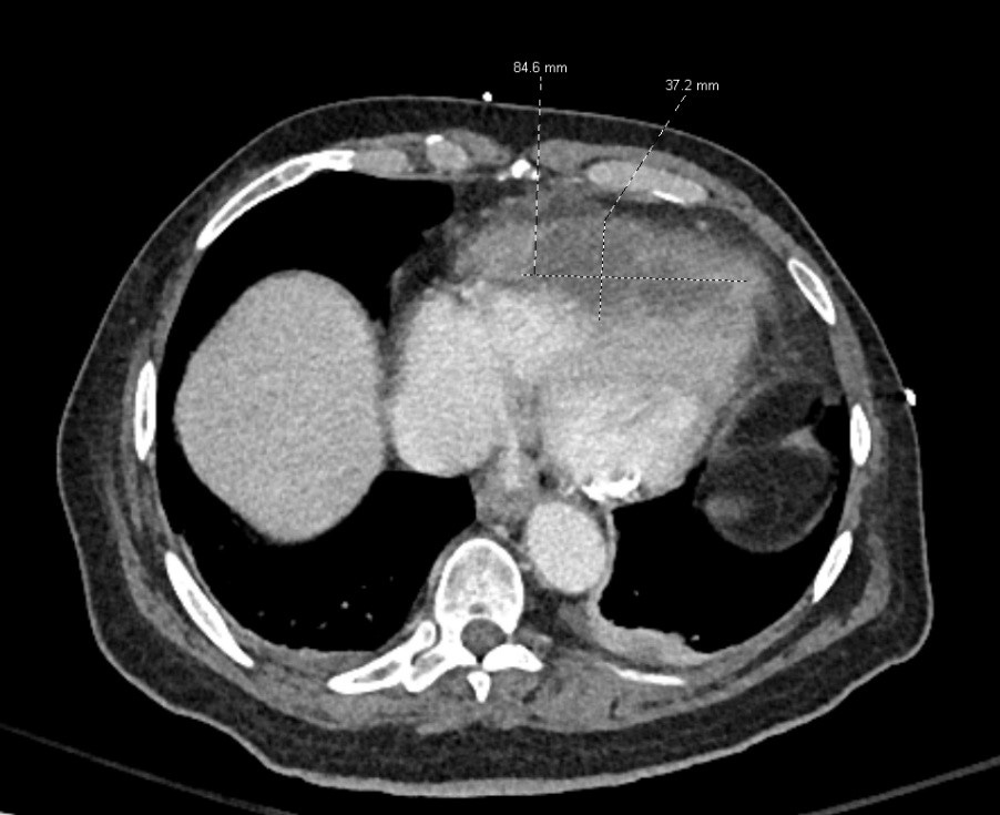
They tell you that they see a complex multiloculated rim enhancing fluid collection, and, based on the images, they’re very concerned for a pericardial abscess.
Interventional cardiology and CT surgery looks over the CT and US images you sent, come right down, and rush him upstairs for emergent drainage.
Recap:
- There are several important differences between a simple pericardial effusion and pericardial abscess on echo
- Abscesses are often thickened and loculated with internal septations and fibrinous debris
- Simple effusions are often circumferential without internal architecture
- Pericardial abscesses can also be localized and difficult to see
- They will occasionally be mistaken for a pericardial cyst or tumor
- If the transthoracic echo is inconclusive or indeterminant and your suspicion is high, consider a transesophageal echo or CT scan
- These patients are often quite sick with multiple other chronic medical problems, such as diabetes or immunosuppression
- Thank you to Aram and The Echo Journal for their help with this interesting case!
Hit me up with any questions,
Lloyd