
Written by Megan Hilbert
Spoon Feed
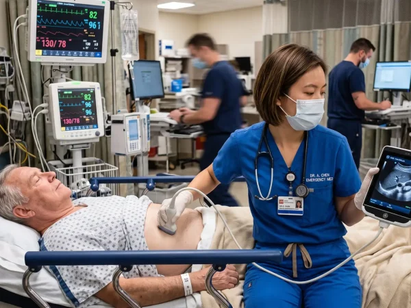
This paper is a great introduction (or reminder) for how to use thoracic ultrasound (TUS) for pleural evaluation.
Quick and dirty TUS
Probe selection:
- High-frequency linear transducer for nearfield assessment (ie: intercostal vessels, parietal pleural thickness, etc.)
- Phased-array transducer for initial assessment, curvilinear transducer for procedural guidance (bigger footprint)
Patient positioning:
- Seated and leaning forward if possible, semirecumbent if not
Evaluate the following:
- Pleural effusion characteristics (echogenicity—anechoic vs. echogenic, septations—present or absent, complex or homogenous)
- Quantitative assessment of volume not widely used in clinical practice
- Inspect pleura for nodularity, thickening >0.3mm (suggests malignancy)
- Always concurrently evaluate the lung for consolidation, compressive atelectasis, interstitial syndrome (b-lines due to varying etiology), presence of mass
- Bedside echocardiogram can complement your findings
Let’s talk procedures:
- When deciding whether to do therapeutic thoracentesis, consider the following: degree of dyspnea, presence of abnormal shape/movement of diaphragm (should not flatten/evert or move cephalad during inspiration if normal), can you pursue medical management instead?
- It can guide the decision to pursue thoracentesis vs. small-bore chest tube placement depending upon characteristics of effusion (more complex likely warrants small-bore chest tube). May need to consider consultation from Thoracic Surgery depending upon degree of complexity.
- Evaluate for post-procedural complications such as pneumothorax or intrapleural bleeding.
Consider use of the following algorithm provided by these authors:

The images included in this paper are also fantastic and warrant a gander to remind yourself of the following specifics: spine sign, curtain sign, sinusoid sign, hematocrit sign, suspended microbubble sign, empyema necessitans, and plume sign.
How will this change my practice?
It is easy to fool ourselves into thinking that lung ultrasound and the evaluation of b-lines and pneumothorax is the only way we can use a probe on the chest. If you feel uncomfortable with TUS, haven’t heard of the terms and descriptions above, or don’t frequently use it in your practice, I highly recommend that you read this paper.
Source
Thoracic Ultrasound in Pleural Effusion: A Bedside Practical Approach Introduction . Chest. 2026 Aug 18:S0012-3692(26)06360-9. doi: 10.1016/j.chest.2026.08.013. Epub ahead of print. PMID: 42612902.