פוסט זה זמין גם ב:
![]() עברית
עברית
Davide Giordano, M.D., and Carmine Pernice, M.D.
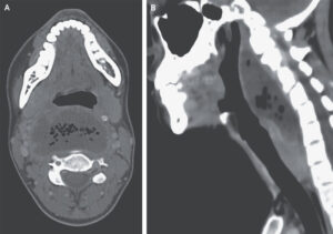
A 26-year-old woman with a history of recurrent tonsillitis presented to the emergency department with a 2-week history of progressive sore throat and a 7-day history of pain when swallowing, shortness of breath, and fever. She reported no recent sexual contact or throat trauma. On physical examination, there was marked swelling of the posterior oropharynx. Laboratory evaluations showed a white-cell count of 19,450 cells per cubic millimeter (reference range, 4000 to 10,000). Computed tomography of the neck showed a collection of fluid posterior to the pharynx measuring 6.2 by 3.4 cm, with air bubbles and a contrast-enhancing rim (Panel A), findings consistent with a retropharyngeal abscess. The fluid collection measured 10 cm on the sagittal axis (Panel B), but there was no mediastinal involvement and no thrombosis of the internal jugular vein. Urgent surgical drainage was performed, and treatment with broad-spectrum antimicrobial agents was initiated intravenously. Fluid cultures grew Fusobacterium necrophorum. Retropharyngeal abscesses can be life-threatening owing to the risks of severe sepsis, airway obstruction, and contiguous spread to the mediastinum. If there is clinical suspicion of retropharyngeal abscess, cross-sectional imaging should be performed. By postoperative day 7, the patient’s symptoms had abated, and she was discharged with oral antimicrobial agents.