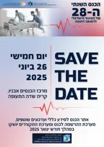
פוסט זה זמין גם ב:
![]() עברית
עברית
Jan Rudolph, MD; Christian Huemmer, PhD; Alexander Preuhs, PhD; Giulia Buizza, PhD; Boj F. Hoppe, MD; Julien Dinkel, MD; Vanessa Koliogiannis, MD; Nicola Fink, MD; Sophia S. Goller, MD; Vincent Schwarze, MD; Nabeel Mansour, MD; Vanessa F. Schmidt, MD; Maximilian Fischer, MD; Maximilian Jörgens, MD; Najib Ben Khaled, MD; Thomas Liebig, MD; Jens Ricke, MD; Johannes Rueckel, MD; and Bastian O. Sabel, MD
Abstract 

Background: Chest radiographs (CXRs) are still of crucial importance in primary diagnostics, but their interpretation poses difficulties at times.
Research question: Can a convolutional neural network-based artificial intelligence (AI) system that interprets CXRs add value in an emergency unit setting?
Study design and methods: A total of 563 CXRs acquired in the emergency unit of a major university hospital were retrospectively assessed twice by three board-certified radiologists, three radiology residents, and three emergency unit-experienced nonradiology residents (NRRs). They used a two-step reading process: (1) without AI support; and (2) with AI support providing additional images with AI overlays. Suspicion of four suspected pathologies (pleural effusion, pneumothorax, consolidations suspicious for pneumonia, and nodules) was reported on a five-point confidence scale. Confidence scores of the board-certified radiologists were converted into four binary reference standards of different sensitivities. Performance by radiology residents and NRRs without AI support/with AI support were statistically compared by using receiver-operating characteristics (ROCs), Youden statistics, and operating point metrics derived from fitted ROC curves.
Results: NRRs could significantly improve performance, sensitivity, and accuracy with AI support in all four pathologies tested. In the most sensitive reference standard (reference standard IV), NRR consensus improved the area under the ROC curve (mean, 95% CI) in the detection of the time-critical pathology pneumothorax from 0.846 (0.785-0.907) without AI support to 0.974 (0.947-1.000) with AI support (P < .001), which represented a gain of 30% in sensitivity and 2% in accuracy (while maintaining an optimized specificity). The most pronounced effect was observed in nodule detection, with NRR with AI support improving sensitivity by 53% and accuracy by 7% (area under the ROC curve without AI support, 0.723 [0.661-0.785]; with AI support, 0.890 [0.848-0.931]; P < .001). Radiology residents had smaller, mostly nonsignificant gains in performance, sensitivity, and accuracy with AI support.
Interpretation: We found that in an emergency unit setting without 24/7 radiology coverage, the presented AI solution features an excellent clinical support tool to nonradiologists, similar to a second reader, and allows for a more accurate primary diagnosis and thus earlier therapy initiation.
Keywords: AI assistance; artificial intelligence; chest radiography; emergency unit.