Written by Clay Smith

This is the latest on how to work up possible acute coronary syndrome in the ED.
Why does this matter?
The AHA released¬†Chest Pain Guidelines¬†in 2021. The ACC thought, вАЬpractical guidance was needed.вАЭ
My troponin is leaking
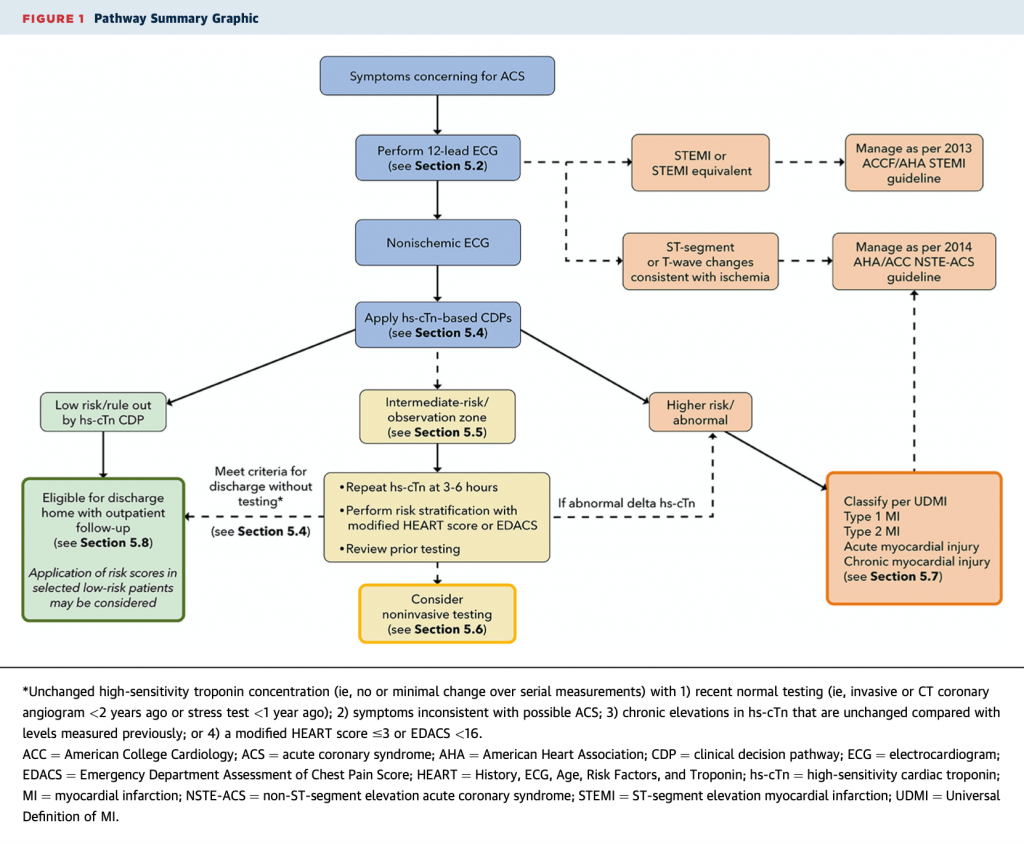
Here is the algorithm and my thoughts below.

Symptoms:¬†We say вАЬchest pain,вАЭ but ask patients about chest¬†discomfort¬†(pressure, tightness, squeezing, heaviness, burning) or non-chest symptoms (pain in arm, neck, back, epigastrium, jaw). Dyspnea, nausea/vomiting, diaphoresis, fatigue, or mental status change could be ACS as well. Pleuritic, positional, or pain at a single small point on the chest wall is unlikely to be ACS. DonвАЩt use вАЬtypicalвАЭ or вАЬatypical;вАЭ use cardiac, possible cardiac, or non-cardiac chest pain.
ECG:¬†STEMI may be subtle in limb leads. Can you detect¬†STEMI equivalents: posterior MI, modified Sgarbossa, DeWinter sign, and hyperacute T waves? For ischemia, would you recognize subtler aVR elevation plus global ST depression (left main disease) or¬†WellenвАЩs Syndrome? Compare with an old ECG when you can.
hs-Tn-based Clinical Decision Pathways (CDP): A hs-Tn approach is recommended first, before other CDPs. Cutoff values for 99th percentile and level of quantification depend on the assay used. The European Society of Cardiology 0/1 and 0/2-hour or High-STEACS rule-outs are recommended. Assay-specific info in the full text is invaluable.
When to use HEART or EDACs:¬†If you use conventional troponin or a patient is intermediate risk with a hs-Tn assay, use HEART or EDACS plus serial conventional troponins at 0 and 3 hours (or repeat hs-Tn at 3-6 hours). If a patient has no change in troponin/hs-Tn (i.e. a 20% increase)¬†AND¬†has had recent normal testing* or unchanged chronic troponin elevation or HEART is вЙ§3 / EDACS <16, consider discharge. If troponin rises, symptoms or ECG worsen вАУ admit. What if patients are intermediate risk at this step? Consider non-invasive testing.
*Recent normal testing is considered an invasive or CT coronary angiogram <2 years without evidence of coronary plaque or a stress test <1 year without ischemia
Non-invasive testing: Use for intermediate risk patients. Consider coronary CTA or other testing: exercise ECG, stress CMR, stress echocardiography, or stress perfusion imaging. Use CCTA if: no known CAD, no known coronary calcification, prior inconclusive non-invasive tests, and no contrast allergy or renal dysfunction. Use other stress tests if: known CAD or calcification, contrast allergy, or renal dysfunction. CCTA may lead to more downstream testing.
So, you have a troponin leak: Is it type 1 (coronary occlusion) or type 2 (supply/demand mismatch)? Consider other causes of myocardial injury: PE, hypertensive emergency, myocarditis, stress cardiomyopathy, contusion, or recent cardiac procedure.
Source
2022 ACC Expert Consensus Decision Pathway on the Evaluation and Disposition of Acute Chest Pain in the Emergency Department: A Report of the American College of Cardiology Solution Set Oversight Committee. J Am Coll Cardiol. 2022 Oct 6;S0735-1097(22)06618-9. doi: 10.1016/j.jacc.2022.08.750. Online ahead of print.


